Virtual Medical Imaging Labs
The goal of this project is to provide Matlab-based lab projects to enhance the biomedical imaging curriculum. Driven by the need from healthcare market, the number of students taking biomedical courses are increasing every year. Considering limited access to the expensive imaging systems, Matlab-based labs are developed to offer students hand-on learning experience without the associated cost and time restrictions. The Virtual Medical Imaging Lab has a MRI simulator and a Ultrasound Imaging Simulator. The Ultrasound Imaging Simulator has two components: 1) B-mode simulator that can be used to demonstrate how imaging parameters and physical anatomy can affect the B-mode images. 2) Simulink module that uncovers the signal processing methods that create B-mode images from raw US signals. The first module allow students to understand the relationships between imaging system and image phantom qualitatively. The second module goes deeper to allow students to tune or develop their own signal processing blocks to extract US image from real data. The MRI simulator lab has three purposes: 1) parameter calculation that tests the students' understanding of the relationship between the input parameters and operating parameters; 2) the acquire module simulates the acquisition process, generating the different gradient form and acquired signals; 3) the show image module shows the experimental data acquired using the listed parameters and the images reconstructed from the acquired data.
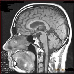
MRI Simulator
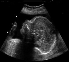
More InfoB-mode Ultrasound Simulator
In this simulator, US signal is modeled as a convolution of point spread function of US scanner with a scatter distribution. The anatomy can be generated from CT images. Shadow can be calculated using reflection coefficient depth and exponential attenuation factor. Deformation due to acoustic speed mismatch is also taken into consideration. To allow a lower computation cost, speckles are simply modeled through Gaussian noises ......
Ultrasound Signal Processing
Signal Processing and Control in SIMULINK provides a programmable platform in SIMULINK that enables the student to visualize the entire signal chain of an ultrasound system. A lot of variability in expert opinion arises from the interpretation of anatomical structures in ultrasound images. Thus it is imperative in knowing the exact processing steps performed in ultrasound system. Staging each processing step as a specialized block breaks ......